
In the ever-evolving world of medical technology, ultrasound imaging isn’t being left behind. The field is bustling with fresh innovations and cutting-edge trends that promise to revolutionize the way we perceive and utilize ultrasound imaging.
From the rise of 3D and 4D imaging to the advent of portable, handheld devices, the ultrasound landscape is shifting rapidly. These advancements are not just enhancing the diagnostic capabilities but also improving patient experience significantly.
Emerging Trends In Ultrasound Imaging
Building on an established foundation of ultrasound technology innovation, advancements in Artificial Intelligence, Point-of-care Ultrasound (POCUS), and the expansion of 3D and 4D imaging techniques constitute the front line of ultrasound imaging trends dominating the contemporary medical landscape.
Role of Artificial Intelligence in Ultrasound Imaging
Artificial Intelligence (AI) presents a groundbreaking approach in the realm of ultrasound imaging. Implementing AI into ultrasound facilitates rapid image analysis helping clinicians make efficient diagnostic decisions. For instance, AI can swiftly identify abnormalities or changes in images, marking them for further scrutiny by medical professionals. AI applications aren’t merely a convenience, they’re a necessity, chipping away at tedious processes, reducing errors, and increasing overall efficiency in diagnostic procedures.
Point-of-care Ultrasound (POCUS) Trend
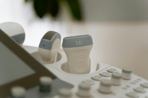
The rising popularity of Point-of-care Ultrasound, abbreviated as POCUS, depicts another significant trend in the sphere of ultrasound imaging. POCUS brings forth a patient-oriented approach, offering diagnostic services directly at the patient’s bedside. It’s not just mobile; it’s agile, granting medical professionals immediate visual information during critical moments. Take, for example, emergency rooms or intensive care units, where POCUS quickly provides visual guidance, optimizing therapeutic procedures and patient outcomes.
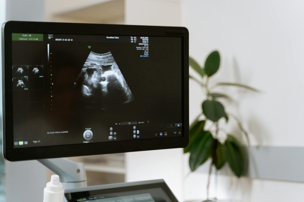
The Rise of 3D and 4D Ultrasound Imaging
Advancing from traditional 2D imaging, the robust growth of 3D and 4D ultrasound imaging techniques elevates diagnostic capabilities. These developing techniques unveil a new dimension of data, creating volumetric, lifelike representations of internal body structures. Specifically, 3D imaging captures still images while 4D captures movement, like a real-time video. The distinction is critical when monitoring dynamic events such as a baby’s movement in a mother’s womb or blood flowing through the heart, enhancing medical diagnostics’ accuracy and effectiveness.
Key Advancements in Ultrasound Imaging Technology
Continued evolution marks the field of ultrasound imaging, bringing forth significant technological advancements. Two critical developments Doppler Ultrasound and Ultrasound Elastography further raise the bar in clinical diagnostics.
Advances in Doppler Ultrasound

Doppler ultrasound, initially perceived as a technique for measuring blood flow and heart conditions, has seen remarkable evolution. The technology’s capabilities have progressed far beyond its original purpose, expanding its applications and refining its functions. For instance, the introduction of color Doppler has improved the visualization of blood flow, serving as a vital tool in investigating vascular disorders.
Particular attention is on Power Doppler or Energy Doppler, which provides sensitivity to low-velocity flow, proving beneficial especially in organs with low blood supply. Incorporation of power Doppler notably enhances the detection and characterization of lesions.
Developments in Ultrasound Elastography
Furthering the boundaries of ultrasound imaging is the advent of elastography, a technique measuring tissue stiffness or elasticity. It offers valuable insights into various medical conditions, like liver fibrosis and breast tumors, where changes in tissue stiffness are involved. In recent years, shear-wave elastography (SWE) emerged as a key improvement. Unlike strain elastography, SWE doesn’t require external compression, making it technically easier and more reproducible, thus increasing its clinical reliability in diagnosing diseases.

Lastly, the concept of point shear wave speed measurements arose, which simplifies the procedure by generating a single elasticity value, enhancing the method’s practicality and convenience. These advancements underline the overall growth and sophistication in ultrasound elastography, assisting clinicians in delivering more precise diagnoses.
Ultrasound imaging’s future looks bright with the rise of AI, POCUS, and expansion in 3D and 4D imaging techniques. These advancements are transforming the healthcare landscape, pushing the boundaries of diagnostic precision, and enhancing patient experiences. As the field continues to evolve, ultrasound imaging’s role in diagnostics is expected to grow, improving patient outcomes and shaping the future of healthcare.